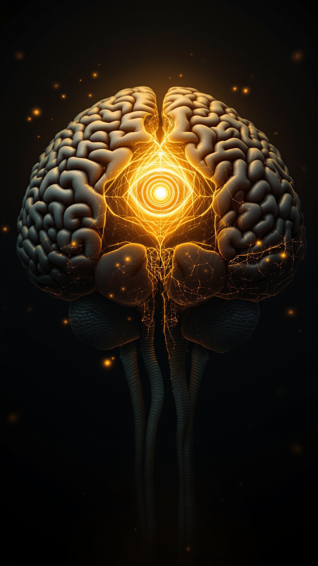
There is a pea-sized gland at the geometric center of your brain. It sits between the two hemispheres, attached to the roof of the third ventricle just above the thalamus. It is the only major brain structure that is not paired — there is exactly one of it, perfectly midline — and the only one that sits outside the blood-brain barrier. Descartes, writing in 1649, called it the seat of the soul. Hindu and Buddhist traditions place the ajna chakra at the same anatomical location and call it the third eye. The Egyptians called it the Eye of Horus. Rick Strassman, writing in 2001, called it the spirit molecule factory. Modern neuroscience calls it the master of circadian biology and the source of melatonin. Each of these descriptions is partly correct. Each is also partly mythology. This is the difference between them.
Where It Sits and What It's Made Of
The pineal gland (epiphysis cerebri, from the Greek for pine cone, after its shape) is approximately 5–8 mm long and weighs roughly 100–150 milligrams in adults. It sits in the epithalamus, dorsal to the third ventricle, attached to the brain via the habenular and posterior commissures. Histologically it is composed primarily of pinealocytes — neuroendocrine cells derived from neuroepithelium — supported by astrocyte-like glial cells and richly vascularised. The gland's vascularisation is exceptional: per gram of tissue it receives a blood flow second only to the kidney. This is mechanistically critical. Because the pineal sits outside the blood-brain barrier and is so heavily perfused, it can rapidly secrete its products into the systemic circulation — which is exactly what a hormone-producing gland needs to do.
The same property makes the pineal unusually exposed to whatever is circulating in the blood. Pharmacological agents, metals, fluoride, and other compounds that the rest of the brain is protected from can accumulate in the pineal. This is the anatomical basis for both the calcification phenomenon and the various toxicological concerns that have grown up around the gland.
Evolutionary Origin: An Eye That Wasn't
The pineal gland's evolutionary backstory is one of the most interesting in vertebrate biology. In primitive vertebrates — lampreys, the tuatara reptile, several lizard species — the pineal complex includes a structure called the parietal eye or third eye that is genuinely photosensitive, possessing a lens, retina-like photoreceptors, and a nerve connection to the brain. The tuatara's parietal eye is so well-developed in young individuals that you can see it on top of the head as a small patch of differentiated skin. It does not produce vision in the conventional sense, but it does detect light and contributes to circadian and seasonal physiology.
Mammals lost direct pineal photoreception over evolutionary time. The modern mammalian pineal is purely endocrine — it does not respond directly to light at all. Instead, light information arrives indirectly: photoreceptors in the retina detect light, signals travel through the retinohypothalamic tract to the suprachiasmatic nucleus (SCN) of the hypothalamus, the SCN signals the superior cervical ganglion, and from there sympathetic nerve fibres reach the pineal and modulate melatonin production. The "third eye" was once literal in your ancestors. It is now a metaphor.
The Master of Time: Melatonin and Circadian Biology
The pineal gland's documented, biochemically proven function is the synthesis of melatonin (N-acetyl-5-methoxytryptamine) from serotonin. The pathway proceeds in two enzymatic steps: arylalkylamine N-acetyltransferase (AANAT) acetylates serotonin to N-acetylserotonin; then hydroxyindole-O-methyltransferase (HIOMT, also called ASMT) methylates the hydroxyl group at the 5-position to produce melatonin. The reactions occur at night. The enzymes are downregulated during daylight. The result is a pronounced circadian rhythm in melatonin output: virtually undetectable during the day, rising rapidly in the early evening, peaking in the middle of the night around 2–3 AM, declining toward dawn.
This circadian melatonin signal is the brain's master timekeeper. It does not generate the circadian rhythm — that is the SCN's job — but it propagates the rhythm throughout the body. Every cell in the body has melatonin receptors (MT1 and MT2), and the nightly melatonin pulse synchronises gene expression, immune function, antioxidant defences, and metabolism across virtually all tissues. The pineal is, functionally, the body's conductor of time.
What Disrupts the Conductor
Modern life disrupts pineal melatonin output more than almost any environmental factor of the last 200 years. Bright light at night — especially short-wavelength blue light from screens — suppresses melatonin synthesis directly. Artificial light at typical residential levels can shift the circadian rhythm by hours within a single night. Chronic shift work has been classified by the IARC as a probable carcinogen (Group 2A) partly through this mechanism: disrupted melatonin signalling impairs nightly tissue repair and antioxidant defence.
The intervention that actually supports pineal function, then, is not a supplement or a "decalcification" diet. It is behavioural circadian hygiene: morning sunlight to the eyes, consistent sleep timing, dim warm light in the evening, blackout-dark sleeping conditions, avoidance of late-night blue light. The gland responds to environmental signal patterns far more than to anything you can buy in a bottle.
Modern light environments are the primary suppressor of pineal melatonin output. Before electric lighting, humans spent ten to fourteen hours per night in functional darkness. The modern urban human spends approximately one hour in true darkness per night. The melatonin amplitude that protects, repairs, and synchronises every cell in the body has been quietly cut in half by lighting choices nobody made deliberately.
OOTW Mushroom Chocolate
Precision-dosed functional mushroom chocolate — engineered for daily ritual, neural support, and sustained clarity. Lab-tested, ceremony-ready.
Shop Mushroom Chocolate →Beyond Sleep: The Neuroprotective and Antioxidant Role
Melatonin is widely misunderstood as merely "the sleep hormone." It is in fact one of the most potent endogenous antioxidants known. Unlike most antioxidants, which become pro-oxidant after they donate an electron, melatonin's metabolic products (cyclic 3-hydroxymelatonin, AFMK, AMK) are themselves antioxidants — creating a "free radical scavenging cascade" in which a single melatonin molecule can neutralise multiple reactive species. It crosses cell membranes freely, accumulates in mitochondria where reactive oxygen species are generated, and protects mitochondrial DNA from oxidative damage.
The neuroprotective implications are significant. In animal models of Alzheimer's disease, melatonin treatment inhibits both β-amyloid deposition and tau hyperphosphorylation — the two pathological hallmarks of the disease. It suppresses neuroinflammation in models of Parkinson's disease, stroke, and traumatic brain injury. Whether exogenous melatonin supplementation translates these effects to humans at clinically meaningful magnitudes is still being established; the dose-response is not simple, and chronic exogenous melatonin can downregulate endogenous receptor sensitivity. But the picture that emerges is of a hormone whose evolutionary role goes far beyond "make you sleepy at night."
The Immune System Connection
Melatonin also modulates immune function. T-helper cells express melatonin receptors. Pineal output peaks coincide with nightly immune surveillance activity. Chronic sleep deprivation — which compresses the melatonin peak — is associated with impaired immune response to vaccination and increased susceptibility to upper respiratory infection. The pineal–melatonin axis sits at the centre of the body's nightly maintenance programme. When it works, you wake up repaired. When it doesn't, the damage accumulates.
The DMT Story: Strassman to Borjigin
The most contested and culturally prominent claim about the pineal gland is that it produces DMT — N,N-dimethyltryptamine, the structurally simple but pharmacologically extraordinary tryptamine that is the active ingredient in ayahuasca, in 5-MeO-DMT's cousin medicines, and in the most reliable peak mystical-experience occasioning agent known. The case for pineal DMT production was made most famously by Rick Strassman in his 2001 book DMT: The Spirit Molecule, where he proposed that endogenous pineal DMT release during birth, near-death experiences, and dream states accounted for the consistent phenomenology of those states across cultures.
Strassman's hypothesis was theoretical. He had not detected DMT in the pineal — he had argued, on indirect biochemical grounds (the enzymes appeared to be present, the precursor tryptamine appeared to be available, the gland's anatomical position suggested it), that the pineal was a plausible candidate for endogenous DMT synthesis. The empirical work to confirm or refute the hypothesis took more than a decade to assemble.
Barker & Borjigin (2013): Direct Detection
The breakthrough came in 2013 from a collaboration between Steven Barker at Louisiana State University and Jimo Borjigin's lab at the University of Michigan. Using liquid chromatography–tandem mass spectrometry on rat pineal microdialysate, the team detected DMT — along with 5-methoxy-DMT and bufotenine — in the pineal in vivo. The concentrations were low (nanomolar range), but they were unambiguous. Strassman's hypothesis had its first empirical confirmation: the mammalian pineal does in fact produce DMT.
Dean & Borjigin (2019): DMT Is Everywhere
The 2013 finding was almost immediately complicated. In 2019, Jon Dean and the Borjigin lab published in Scientific Reports (PMID:31249368) a landmark study using in situ hybridisation to localise the two enzymes required for DMT synthesis: aromatic L-amino acid decarboxylase (AADC) and indolethylamine N-methyltransferase (INMT). Both enzymes were expressed not only in the pineal but extensively throughout the cortex and the hippocampus — brain regions central to perception, memory, and higher cognition. The implication was decisive: the pineal is not the unique site of endogenous DMT synthesis. The biochemical machinery for DMT production is distributed across the mammalian brain.
This is the empirical fact that has yet to fully reshape the popular pineal-DMT narrative. The pineal can produce DMT. So can your cortex. The "spirit molecule factory" is not a single small gland — it is, potentially, large portions of your entire brain.
Borjigin (2023): The Dying Brain
The most recent and most provocative work from the Borjigin lab examined DMT levels in dying rat brains. Using microdialysis sampling during induced cardiac arrest, the team found that DMT levels surged in some animals during the period coinciding with the gamma-wave electroencephalographic signature also seen in dying human brains. Critically, the DMT surge was preserved in animals that had previously undergone pinealectomy — surgical removal of the pineal gland. The DMT did not require the pineal to produce it. The cortex, the hippocampus, or some other distributed site was sufficient.
The same year, the Borjigin lab published their landmark study of dying human brains, documenting a brief but extraordinary surge of high-frequency gamma oscillations and cross-region functional connectivity in the seconds following cardiac arrest in two patients (PMID: see PNAS, May 2023). They were careful in their interpretation: the data is consistent with the long-reported phenomenology of near-death experiences, but it does not prove that DMT is the cause, and the relationship between the gamma surge and any subjective experience remains untested.
The verdict on endogenous DMT: it exists, it is produced by the mammalian brain including the pineal, but the pineal is not the unique source. The Strassman hypothesis is partly vindicated and partly refined. The most extraordinary claims — that pineal DMT release is the mechanism of dream consciousness, near-death experiences, or the felt sense of mystical unity — remain hypotheses awaiting decisive evidence.
OOTW Spirit Guide
Set. Setting. Dose. Integration. The questions you can't bring to your doctor — answered by an AI grounded in every peer-reviewed paper, protocol, and ceremony manual. Private, sober, always there.
Talk to the Spirit Guide →The Calcification Question
The pineal gland accumulates calcium deposits called corpora arenacea — literally "sandy bodies," also called brain sand or acervuli. The deposits begin to form in childhood and become near-universal by middle age. They are visible on MRI and on plain X-rays of the skull. Functionally, the calcification is sometimes described as inert anatomical curiosity; in popular discourse it has acquired a much larger significance as a marker of pineal "blockage" and a target for various decalcification protocols.
What the Evidence Actually Shows
Pineal calcification is correlated with age — virtually everyone over 60 has detectable corpora arenacea, and the prevalence increases monotonically from childhood through senescence. Whether the calcification meaningfully impairs melatonin output is less clear. Several studies have found correlations between pineal calcification and lower nocturnal melatonin levels, and between calcification and disrupted sleep in older adults. Other studies have not replicated these findings. The mainstream view in pineal physiology — articulated in the comprehensive 2018 Molecules review by Tan, Manchester, and Reiter (PMC6017004) — is that calcification likely reduces functional pineal tissue volume and therefore reduces maximum melatonin output, but that the relationship is not as deterministic as popular accounts suggest.
The Fluoride Question
Jennifer Luke's 1997 doctoral thesis at the University of Surrey, later published in 2001, was the foundational documentation that fluoride accumulates preferentially in the pineal gland. Because the gland is heavily vascularised and sits outside the blood-brain barrier, fluoride from systemic circulation deposits in pineal corpora arenacea at concentrations significantly higher than in other soft tissues. Luke's measurements found fluoride concentrations in pineal calcifications comparable to those in dental enamel.
What this means for pineal function is contested. Luke proposed that pineal fluoride accumulation could impair melatonin synthesis; subsequent studies have produced mixed results. The 2024 National Toxicology Program systematic review of fluoride and neurodevelopment found inverse associations between fluoride exposure and IQ in children, but the exposure levels in those studies were almost exclusively above 1.5 mg/L — substantially above the U.S. drinking water standard of 0.7 mg/L. The available evidence does not support strong claims that typical municipal fluoridation at U.S. levels significantly suppresses human pineal function. It also does not refute that possibility. The data is genuinely incomplete.
Should You "Decalcify"?
The popular decalcification protocols — boron, vitamin K2, iodine, raw apple cider vinegar, distilled water, various herbs — are not supported by evidence as treatments that reverse existing pineal calcification. Once corpora arenacea form, they generally remain. The mechanisms claimed for these interventions are not well-established. This is not a counsel of despair: the pineal gland responds vigorously to behavioural circadian hygiene, light exposure patterns, and sleep timing. The evidence-based path to good pineal function is profoundly boring: see the sun in the morning, dim the lights at night, sleep dark, sleep consistently. The gland that conducts your circadian symphony rewards exactly the kind of attention you would give a sensitive musical instrument.
The Third Eye: Symbol, Memory, Metaphor
The third-eye mythology is older than recorded history. Hindu tradition places the ajna chakra at the forehead between the eyebrows and identifies it with intuition, perception beyond sensory limits, and the site of inner sight. Egyptian iconography depicts the Eye of Horus at the same anatomical region. Buddhist traditions describe the cultivation of the divya chakshu, the divine eye, through advanced meditative attainment. The pineal gland's geometric position in the brain — midline, central, unpaired — has made it the natural anatomical correlate for these symbolic frameworks across cultures that had no direct contact with each other.
What the modern scientific picture suggests is that the convergence is not mystical coincidence and not literal anatomy. It is something more interesting: contemplative practices across traditions reliably produce changes in attention, prefrontal activity, default-mode network engagement, and subjective experience. The location at which these changes are felt — between the eyebrows, central, forward — is consistent with the somatic attention that meditation practitioners direct to that region. The pineal gland sits roughly behind that point, but the experiential phenomenon is mediated by cortical and subcortical attention networks, not by the gland itself. The third eye is real as an experience. It is not located in the pineal in the literal sense. But the traditions that pointed at the middle of the forehead and said "there" were pointing at something that the modern neuroscience of attention and meditation now studies systematically.
Descartes Revisited
René Descartes, in Les Passions de l'Âme (1649), proposed that the pineal gland was the seat of the rational soul — the unique point at which the immaterial mind interacted with the material body. His choice was not arbitrary. He reasoned that the seat of consciousness should be unpaired (it is), midline (it is), and accessible to inputs from both hemispheres (the pineal does receive from both via the habenular commissure). He was wrong about the specifics — consciousness is not localised in a single small gland; it emerges from distributed cortical networks — but he was attempting to think rigorously about a real problem. The 21st-century scientific consensus has moved past Descartes's specific claim but has not yet solved the harder problem he was after: how does the integrated felt experience of being a conscious self arise from the distributed activity of billions of neurons?
The Verdict: What's Real, What's Myth, What's Unknown
The pineal gland is one of the most extraordinary organs in the body. It is the master conductor of circadian biology. It produces a hormone (melatonin) that protects, repairs, and synchronises every cell in the body each night. It does produce endogenous DMT, alongside cortical and hippocampal sources, in concentrations and conditions that remain incompletely understood. It calcifies with age. It accumulates fluoride. The popular mythologies surrounding it — Descartes's soul-seat, Strassman's spirit molecule factory, the New Age decalcification protocols — are not entirely wrong. They are inexact, sometimes substantially. The truth is more interesting than the myth, but it is also less narratively satisfying.
The actionable conclusions are mundane. Honor your circadian rhythm. See sunlight in the morning. Dim the lights at night. Sleep in darkness, at consistent times. Do not chase decalcification protocols that lack evidence. Understand that endogenous DMT exists in your brain and that its function is one of the genuinely open questions of contemporary neuroscience. Sit with the fact that the pineal gland is a real organ that does real work, and that the mythological frameworks built around it across the centuries were pointing at something — even when they pointed inexactly — that the science is now only beginning to characterise.
Descartes had it half right. Strassman had it half right. The Vedic tradition had it half right. The science, three and a half centuries after Descartes and a quarter century after Strassman, also has it half right. The pineal gland is what it is: a small endocrine organ at the center of your skull, producing the hormone that conducts the body's nightly orchestra, occasionally producing the most powerful psychedelic compound known, and reminding us that some of the most interesting territory in human biology lies not at the frontiers we expected but at the dead center of what we already had names for.
Ceremony-Ready Mushroom ConfectioneriesBring the Science Home
Every article here is the why. OOTW's ceremony-ready mushroom confectioneries are the how — precision-crafted to carry the medicine into your daily practice.
Shop Mushroom Chocolate →